RS
Written for patients of
Dr. Rajesh Soni

Laser, endourological and kidney transplantation care led by Dr. Rajesh Soni at Soni Hospital, Ramdaspeth.

The conditions patients most often see Dr. Soni for at Soni Hospital, Ramdaspeth.
Holmium laser, flexible ureteroscopy and urodynamic evaluation — treatments carried out without an external cut wherever possible.
Kidney stones broken from inside with a flexible scope and laser, or through a small keyhole for larger stones.
Laser and endoscopic treatment for an enlarged prostate with minimal bleeding.
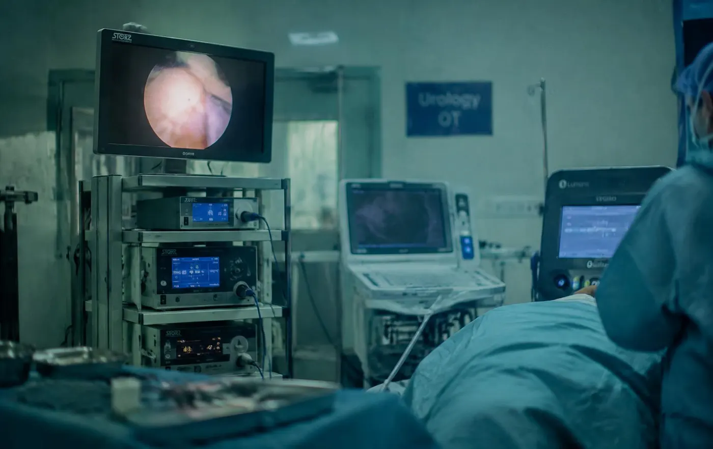 ● Soni Hospital OT
Trained at Muljibhai Patel Urological Hospital, Nadiad and in advanced endourology in Vienna, Dr. Soni founded Soni Hospital in 1998 — a superspeciality centre for laser urosurgery in Central India.
Meet the DoctorFilter by area to find the condition you want to read about.
RIRS, Mini PERC, PCNL and holmium laser lithotripsy for stones of every size.
Flexible ureteroscopic laser surgery — no cut, no tract.
Assessment and treatment of difficult urination in men — from medication to HoLEP.
Holmium laser enucleation of the prostate — size-independent with minimal bleeding.
Time-tested endoscopic prostate resection for moderate gland enlargement.
Endoscopic cystolithotripsy and laser fragmentation for bladder calculi.
Comprehensive surgical management of kidney, prostate, bladder, and testicular cancers.
Living donor renal transplantation and vascular access surgery for end-stage renal disease.
Microsurgical varicocelectomy, TESA/PESA, and comprehensive male reproductive evaluation.
Confidential diagnostic evaluation, medical therapies, and penile implant surgery.
Investigation and targeted management of complicated and recurrent UTIs in men and women.
Optical urethrotomy, anastomotic and buccal mucosa graft urethroplasty.
Hypospadias, undescended testis, VUR and female stress urinary incontinence (TVT/TOT).
Please bring any past ultrasound, CT scan or urine culture reports. Cashless insurance support is available for major insurers and TPAs.
0712-2427103 · Monday – Friday 10:00 AM – 3:00 PM · 6:00 PM – 7:30 PM
Direct Call: 0712-2427103 →Can't find the answer? Call us at 0712-2427103.
A urologist diagnoses and treats disorders of the kidneys, ureters, bladder and urethra in men and women, as well as the male reproductive system — from kidney stones and prostate enlargement to cancers, infertility and reconstructive problems.
Call the appointment desk at 0712-2427103. OPD runs Monday to Friday 10 AM – 3 PM and 6 PM – 7:30 PM, and Saturday 10 AM – 3 PM.
Please bring any past ultrasound, CT scan or urine culture reports, along with a list of current medicines.
Soni Hospital provides cashless claim support for major insurance companies and TPAs. The TPA desk can be reached at +91-9975552181.
Shar-Hari Complex, Central Bazar Road, Ramdaspeth, Nagpur - 440010, Maharashtra, India. Landmark: Directly opposite Somalwar High School.
Clear advice on symptoms, treatment and recovery.
 Symptoms
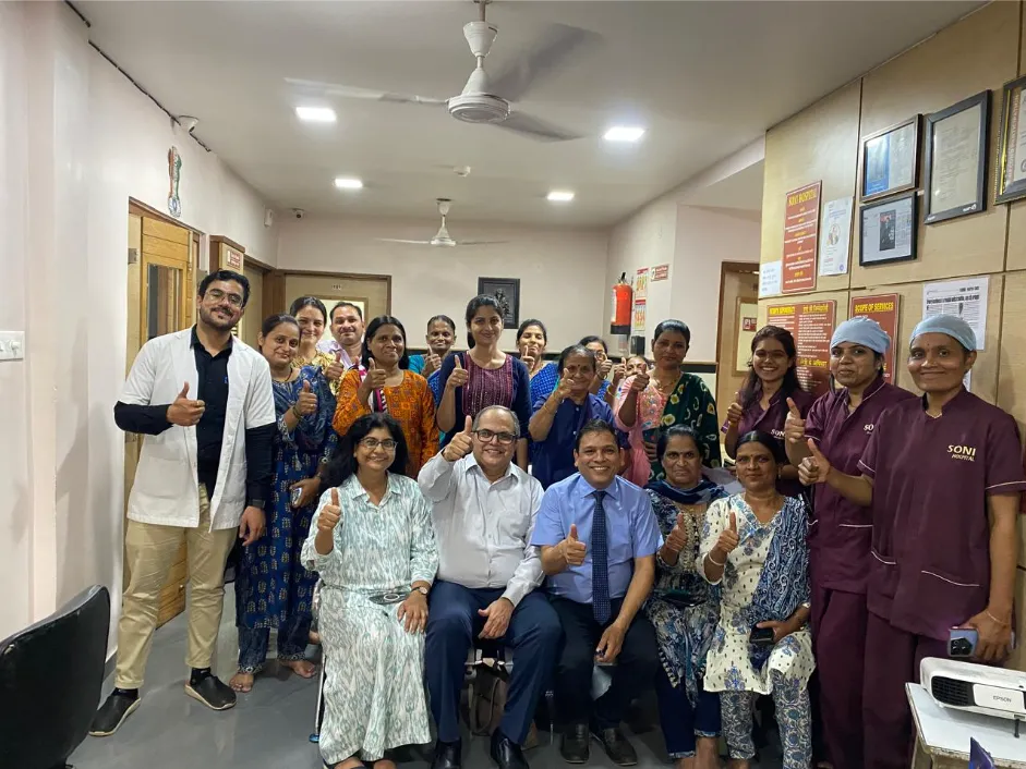 Your visit
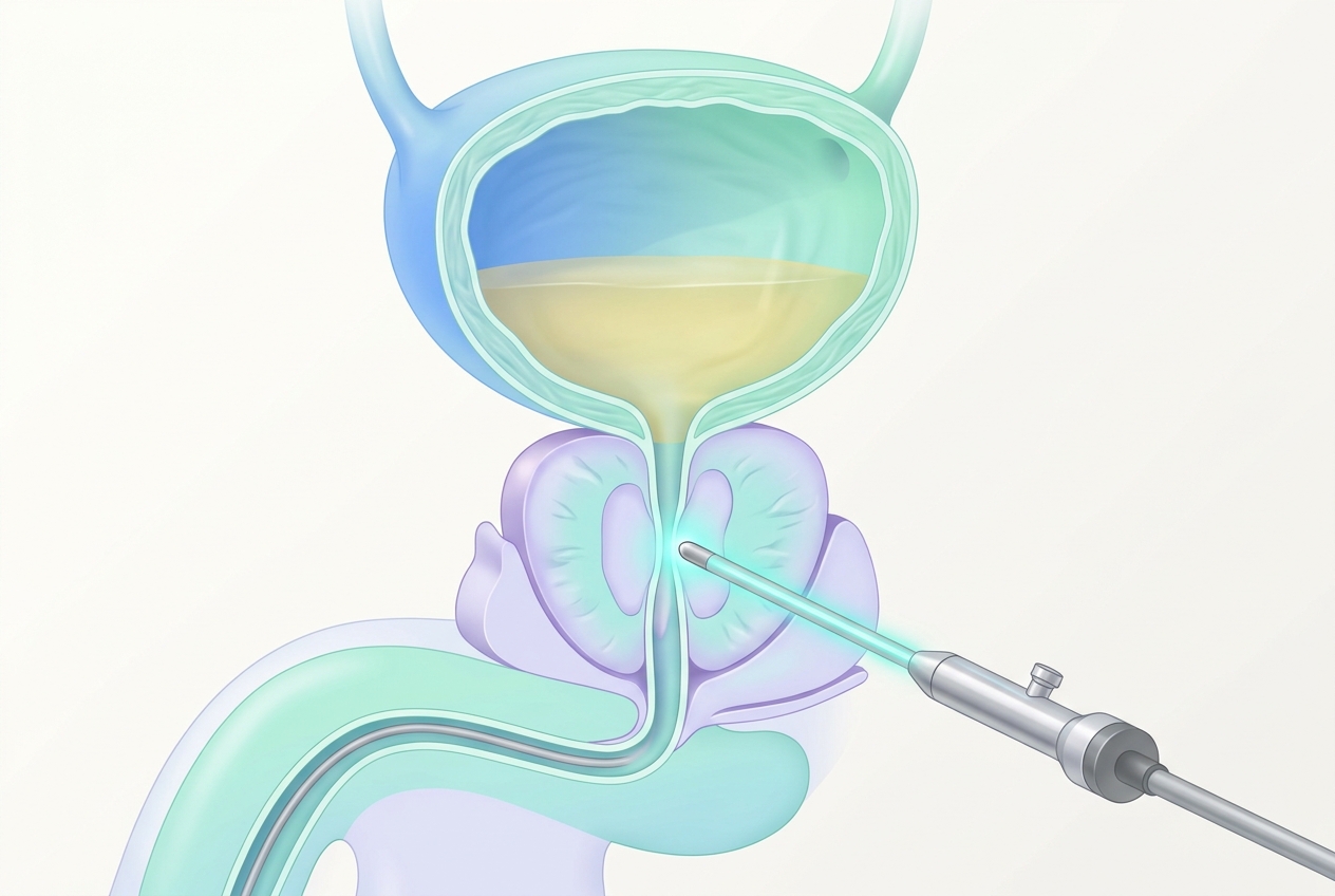 Prostate
Discovering blood in your urine—clinically termed hematuria—is one of the most alarming symptoms a patient can observe. The urine may appear faintly pink, deep amber, bright red, or dark brownish like cola. In other instances, blood is not visible to the naked eye at all, but is discovered incidentally during a routine health check-up or pre-employment urine microscopy. Regardless of whether the discoloration is glaringly visible or microscopic, blood in the urinary tract is never normal and should never be ignored.
At Soni Hospital in Ramdaspeth, Nagpur, Senior Consultant Urologist Dr. Rajesh Soni treats hundreds of patients with hematuria each year. One of the most dangerous clinical pitfalls is the tendency for hematuria to be intermittent: bleeding may occur for a single day, disappear completely for weeks or months, and give the patient a false sense of security. This comprehensive medical guide explains the anatomical origins of urinary bleeding, outlines essential diagnostic investigations, and explains why painless bleeding demands immediate expert evaluation.
In clinical urology, hematuria is categorized into two distinct presentations based on visibility:
Gross hematuria means blood is clearly visible to the naked eye. It takes as little as 1 milliliter of whole blood in one liter of urine to produce a distinct reddish hue. Patients may notice clots resembling small earthworms or stringy strands, which indicate significant bleeding from the upper urinary tract or bladder neck. Gross hematuria carries a 15% to 25% probability of underlying urological malignancy in adult patients, making urgent investigation mandatory.
Microscopic hematuria occurs when the urine appears completely clear and normal in color, but formal laboratory centrifuge examination reveals three or more red blood cells (RBCs) per high-power field (HPF) on microscopic analysis. While often detected during routine physicals, asymptomatic microscopic hematuria still mandates risk stratification and imaging, particularly in individuals with smoking history or occupational exposure to chemicals.
The most important clinical distinction in evaluating hematuria is the presence or absence of accompanying pain:
Bladder tumors frequently bleed intermittently. Fragile new blood vessels on the surface of a papillary tumor may rupture, shed blood for 24 to 48 hours, and then form a temporary clot, restoring clear urine for several months. Clear urine following an episode of bleeding does NOT mean the underlying condition has resolved. Delaying evaluation allows early superficial tumors to invade the deep bladder muscle, transforming a simple endoscopic procedure into complex radical surgery.
The urinary tract extends from the microvasculature of the renal glomeruli down to the external urethral meatus. Bleeding can originate at any point along this anatomical conduit:
| Anatomical Level | Most Common Pathologies | Associated Clinical Clues |
|---|---|---|
| Kidneys (Upper Tract) | Renal cell carcinoma, kidney calculi, glomerulonephritis, polycystic kidney disease, renal trauma. | Flank mass, dull loin ache, protein in urine, red blood cell casts. |
| Ureters | Ureteric calculi, upper tract urothelial carcinoma, stricture disease. | Acute spasmodic renal colic pain, worm-like stringy blood clots. |
| Urinary Bladder | Urothelial bladder carcinoma, radiation cystitis, severe bacterial UTI, bladder calculi. | Painless bleeding, frequency, sudden urgency, necrotic tissue fragments. |
| Prostate Gland (Men) | Benign prostatic hyperplasia (BPH), prostate adenocarcinoma, acute prostatitis. | Initial hematuria (blood at start of stream), weak flow, nocturia, elevated PSA. |
| Urethra | Urethritis, urethral stricture, traumatic catheter insertion, foreign body. | Terminal hematuria, blood spotting on undergarments independent of voiding. |

At Soni Hospital, Dr. Rajesh Soni adheres to evidence-based international clinical guidelines to provide a comprehensive, systematic hematuria workup:
A fresh urine specimen is analyzed to confirm true erythrocyte presence (ruling out pseudohematuria from beetroot, rifampicin, or myoglobin), check for active bacterial infection, and perform urine cytology. In cytology, specialized cytopathologists examine exfoliated cells under high magnification to detect malignant transitional cells shed from the urinary lining.
High-resolution sonography provides an immediate, non-invasive assessment of renal parenchymal thickness, hydronephrosis, calculus shadowing, bladder wall thickening, and intravesical exophytic masses. It also measures prostate volume and post-void residual urine volume.
CT Urography is the imaging gold standard for evaluating the kidneys and ureters. Performed with intravenous contrast across unenhanced, nephrographic, and excretory phases, CTU provides exquisite 3D anatomical visualization of the renal parenchyma, detecting tiny renal parenchymal tumors, caliceal abnormalities, and urothelial filling defects in the ureters.
Neither ultrasound nor CT scan can reliably detect small, flat carcinoma-in-situ (CIS) or mucosal lesions under 5 millimeters inside the bladder. Therefore, flexible cystoscopy is an indispensable part of the hematuria workup. Performed using an ultra-fine, flexible fiberoptic scope lubricated with local anesthetic gel, the urologist directly inspects every square millimeter of the bladder mucosa and urethral lining on a high-definition monitor. The procedure takes less than 5 minutes and is virtually painless.
Blood in the urine is not a diagnosis; it is an urgent symptom indicating pathology somewhere along the urinary tract. While common causes such as urinary tract infections and kidney stones are easily treatable, the potential presence of bladder or kidney tumors demands swift, meticulous medical exclusion. If you observe pink, red, or dark urine, contact Dr. Rajesh Soni at Soni Hospital, Ramdaspeth, Nagpur immediately for prompt diagnostic evaluation and peace of mind.
Shar-Hari Complex, Central Bazar Road, Ramdaspeth, Nagpur - 440010, Maharashtra, India · Landmark: Directly opposite Somalwar High School
From the first consultation to follow-up after surgery.